Varicocele Embolisation
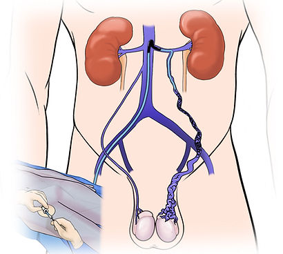
A varicocele is dilation of a vein plexus (group) in the scrotum due to reflux (reversed) flow from typically the left renal (kidney) vein through the gonadal (testicular) vein into the scrotum.
Varicocele
A varicocele is dilation of a vein plexus (group) in the scrotum due to reflux (reversed) flow from typically the left renal (kidney) vein through the gonadal (testicular) vein into the scrotum. This occurs in up to 15% of the male population and is often asymptomatic. Symptoms include dull pain or ache that is commonly worsened by long periods of standing and relieved by lying down. The scrotum may also have a "bag of worms" appearance on examination. Varicocele can also caused decreased fertility, and treatment likely improves fertility chances.
Varicocele Embolisation
Varicocele embolisation refers to the minimally invasive procedure performed by an Interventional Radiologist (IR) to treat the swollen veins in the scrotum. The procedure works by occluding (blocking) the refluxing gonadal (testicular) vein from inside the vein, which prevents the reverse flow from continuing to cause swollen veins in the scrotum. The venous blood flow from the scrotum then diverts to a normal (competent) outflow pathway. Varicocele embolisation is performed as a "pin-hole" day-case operation and is an alternative to major surgery.
Effectiveness
-
90% of men will have improved symptoms following varicocele embolisation
-
Most men (60 to 80%) will have improved sperm count and quality
-
Up to 50% of couples with male-factor infertility achieve successful pregnancies following varicocele embolisation
-
Up to 10% of men will experience recurrence of the varicocele after embolisation, typically several years later, which can be treated with repeat embolisation or surgery
Should I undergo embolisation or microsurgical resection?
Overall, embolisation and microsurgery are both strongly evidence-based procedures for the treatment of varicocele, with similar rates of effectiveness and pregnancy. Advantages of embolisation include a lower rate of complications including hydrocele, however, there may be higher recurrence rates with embolisation compared to microsurgery. The decision of which treatment is best is an individual decision that you should make in consultation with your GP, Interventional Radiologist and Urologist.
Procedure
-
A specialist interventional radiologist will first consult and examine you in the outpatient clinic to assess your suitability for varicocele embolisation. If you are not suitable for embolisation, your Interventional Radiologist (IR) will discuss this with you and arrange referral to another specialist as appropriate.
-
Embolisation is performed under sedation and local anaesthetic, with passage of a small catheter tube into a vein in your neck or groin. The tube is then navigated into the gonadal vein and coils or liquid medication injected to seal the refluxing (reverse flow) vein.
-
You will be required to rest in bed either sitting up or lying down for up to 2 hours post-operatively, which is to minimise bruising.
-
Most patients experience some pain, typically a mild-moderate dull ache, after the procedure, which is treated with pain medications
-
The procedure is performed as a day-case, and you will be able to go home the next day, returning to usual activities including work within 1 to 2 days
-
The vein swelling in the scrotum will gradually shrink down over the weeks following embolisation
Complications
The overall risk of complications is very low (<1%), but these can include:
-
Groin or neck bruising
-
Pelvic or lower back pain (dull ache)
-
Coil migration
Specialist Expertise
Interventional Radiologists (IRs) are specialty trained in the care of patients undergoing varicocele embolisation, including the pre-procedural assessment, operation and post-procedural care. Interventional Radiologists (IRs) are extensively experienced in embolisation procedures as they regularly perform embolisation in many areas of the body, including the lung, liver, spleen, kidney and pelvis.
Preparation and Referral
If you would like to consult with an Interventional Radiologist about varicocele embolisation, please send a referral for consultation, including the following
-
Ultrasound and other imaging results
-
Records of consultations with other doctors and specialists
Public eligible patients can undergo the treatment with no out-of-pocket expense in the public system. Varicocele embolisation is performed at most of our public and private hospital locations in Melbourne, Victoria, Australia.
Varicocele embolisation content by Dr Matthew Lukies.